Peptidoglycan
Mesh-like macromolecule giving bacterial cell walls strength and shape.

Yikrazuul · Public domain
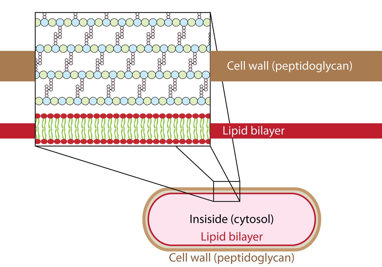
Peptidoglycan, also known as murein or mucopeptide, is a unique large macromolecule and polysaccharide consisting of sugars and amino acids that forms a mesh-like layer (sacculus) surrounding the bacterial cytoplasmic membrane. It serves a structural role in the bacterial cell wall, giving structural strength and counteracting the osmotic pressure of the cytoplasm, and is critical for maintaining cell form and withstanding high osmotic pressures.
- composition
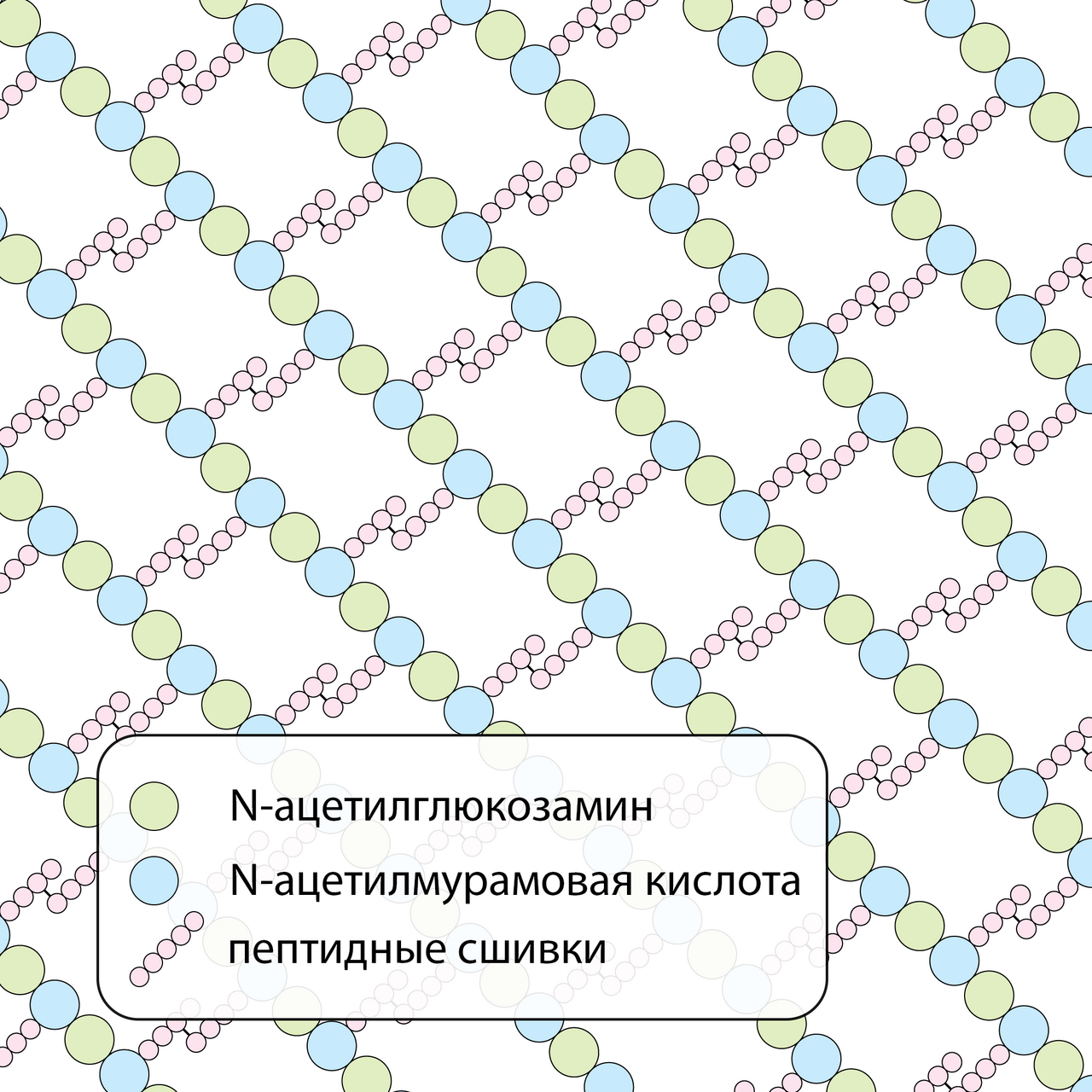
- Alternating residues of β-(1,4) linked N-acetylglucosamine (NAG) and N-acetylmuramic acid (NAM) with oligopeptide chains
- location
- Bacterial cell wall, surrounding the cytoplasmic membrane
- function
- Structural support, counteracting osmotic pressure, maintaining cell shape
- gram-positive_thickness
- 20 to 80 nanometers
- gram-negative_thickness
- 7 to 8 nanometers
- gram-positive_dry_weight
- 40 to 90% of cell wall
- gram-negative_dry_weight
- About 10% of cell wall
Lore & Background
The peptidoglycan layer is substantially thicker in gram-positive bacteria (20 to 80 nanometers) than in gram-negative bacteria (7 to 8 nanometers), and its high levels are the primary determinant of gram-positive characterization. Peptidoglycan is involved in binary fission during bacterial cell reproduction, and its hydrolysis and synthesis must occur for cells to grow and multiply, carried out in three stages: clipping of current material, insertion of new material, and re-crosslinking. The biosynthesis of peptidoglycan monomers occurs in the cytosol, then attached to a membrane carrier bactoprenol, which transports them across the cell membrane for insertion into existing peptidoglycan.
Reader's Guide
Peptidoglycan is fundamental to bacterial biology, providing the structural integrity that allows bacteria to survive and colonize virtually all habitats of the geosphere and hydrosphere. The peptidoglycan layer protects the cell from lysis caused by turgor pressure and maintains cell shape throughout life. The invention of rigid peptidoglycan cell walls in bacteria was probably the prerequisite for their survival and extensive radiation. Peptidoglycan is also a key target for antibiotics such as penicillin, which inhibit the DD-transpeptidase (penicillin-binding protein) involved in cross-linking. Notably, archaea do not contain peptidoglycan; some contain pseudopeptidoglycan instead. The recognition of peptidoglycan by the immune system is an evolutionarily conserved process, with modifications in sugar polymers, cross-linking, or amino acid substitutions increasing diversity.
Did You Know?
- Peptidoglycan is one of the most important sources of D-amino acids in nature.
- Particles of approximately 2 nm can pass through the peptidoglycan in gram-positive bacteria, but gram-negative bacteria have smaller pores that typically allow passage only up to about 1 nm.
- L-form bacteria can proliferate by a variety of mechanisms including budding, blebbing, and tubulation; mycoplasmas typically reproduce by budding, not binary fission.
- The enzyme DD-transpeptidase, which crosslinks glycan chains, is also known as the penicillin-binding protein.
Molecular Architecture and Crystal Lattice
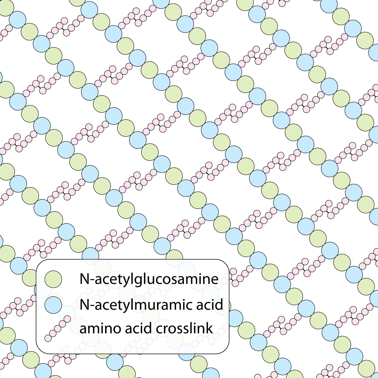
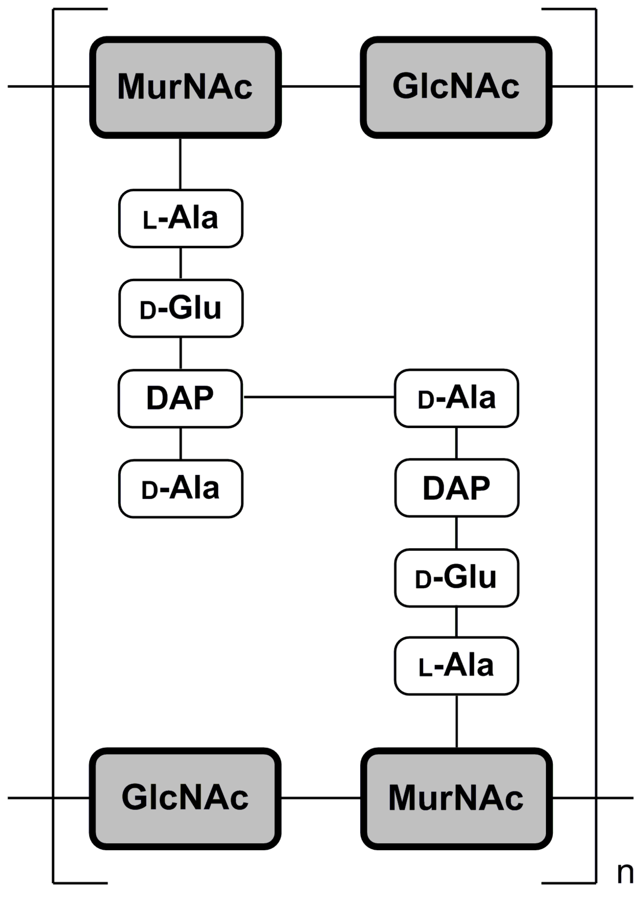
Peptidoglycan is organized as a crystal lattice built from linear polysaccharide chains in which two amino sugars—N-acetylglucosamine and N-acetylmuramic acid—alternate in a repeating pattern held together by β-(1,4)-glycosidic bonds. Every muramic acid residue carries a short oligopeptide tail of three to five amino acids, and the precise composition of that tail differs by species. In the gram-negative bacterium Escherichia coli, the chain includes L-alanine, D-glutamic acid, meso-diaminopimelic acid, and D-alanine, whereas the gram-positive Staphylococcus aureus inserts a five-glycine interbridge between its tetrapeptide units. The enzyme DD-transpeptidase forges covalent bridges between peptide tails on adjacent sugar strands, converting the flat linear chains into a rigid three-dimensional mesh. This cross-linked architecture is what gives the layer its remarkable tensile strength. Peptidoglycan also stands out as one of nature's principal reservoirs of D-amino acids, a stereochemical feature uncommon outside the bacterial world.
Structural Function and Evolutionary Legacy
The peptidoglycan sacculus encloses the cytoplasmic membrane and acts as a pressure-resistant shell that prevents the cell from bursting under the turgor pressure generated by its own cytoplasm. Because the layer is continuously remodeled—old material is clipped away, new monomers are inserted, and existing strands are re-crosslinked to the newcomers—a rod-shaped bacterium remains a rod and a coccus remains a coccus for its entire lifespan. During binary fission, freshly synthesized septal material reshapes into a hemispherical wall that becomes the boundary of each daughter cell. Organisms that completely lack peptidoglycan, such as mycoplasmas and L-form bacteria, cannot divide by this mechanism and instead reproduce through budding. Looking further back in evolutionary time, the emergence of rigid murein walls in the domain Bacteria is widely regarded as the key innovation that allowed bacteria to survive, radiate, and colonize virtually every habitat on Earth, from the geosphere to the hydrosphere. In the earliest stages of life, the development of protective boundaries—membranes and walls—was essential for the transition from molecular assemblies to true cells.
Gram Classification and Diagnostic Significance
The thickness and abundance of the peptidoglycan layer are the defining features that separate gram-positive from gram-negative bacteria. In gram-positive strains the layer measures twenty to eighty nanometers and can account for forty to ninety percent of the cell wall's dry weight, depending on growth pH. Gram-negative organisms, by contrast, possess a much thinner shell of only seven to eight nanometers, contributing roughly ten percent of their wall mass. This stark difference in peptidoglycan content is the primary basis on which bacteria are classified as gram-positive. In those organisms the layer also plays important roles in surface attachment and in serotyping. Despite these differences, particles as small as two nanometers can traverse the peptidoglycan in both groups. The procedure uses crystal violet followed by safranin; gram-positive cells retain the purple dye while gram-negative cells appear pink.
Biosynthesis and Continuous Turnover
Peptidoglycan monomers are assembled in the cytosol and then handed off to a lipid carrier called bactoprenol, which ferries them across the cytoplasmic membrane so they can be incorporated into the existing wall. The first synthetic step involves the enzyme GlmS, which transfers an amino group from glutamine to fructose 6-phosphate, yielding glucosamine-6-phosphate. Next, GlmM acetylates that amino group using acetyl CoA to produce N-acetylglucosamine-6-phosphate. The bifunctional enzyme GlmU then isomerizes the phosphate to the 1-position and, in a second catalytic role, activates the sugar by coupling it to uridine triphosphate, releasing inorganic pyrophosphate and generating UDP-N-acetylglucosamine. A subsequent reaction, catalyzed by a Mur enzyme, appends a lactyl group derived from phosphoenolpyruvate to convert the UDP-NAG into UDP-N-acetylmuramic acid. Because bacterial growth demands constant wall expansion, hydrolysis of old peptidoglycan and synthesis of new material proceed in a tightly coupled cycle of clipping, insertion, and re-crosslinking.
Gallery

Frequently Asked Questions
What is Peptidoglycan?
Peptidoglycan (also called murein or mucopeptide) is a giant macromolecule built from alternating sugar and amino-acid units that assembles into a mesh-like shell, the sacculus, wrapped around the bacterial cytoplasmic membrane. It is, in short, the structural backbone of the bacterial cell wall.
What does Peptidoglycan actually do for a bacterium?
It supplies the mechanical rigidity that prevents the cell from swelling and bursting under internal osmotic pressure. Without this sacculus, a bacterium would lose its defined shape and simply lyse.
How thick is Peptidoglycan in gram-positive versus gram-negative bacteria?
In gram-positive organisms the layer spans roughly 20–80 nm and can account for 40–90 % of the wall's dry weight, while in gram-negative organisms it is a comparatively thin 7–8 nm sheet. This thickness disparity is the structural basis behind the classic Gram-stain color split.
What are the building blocks of Peptidoglycan?
The sugar backbone alternates between N-acetylglucosamine (NAG) and N-acetylmuramic acid (NAM) linked by β-(1,4) glycosidic bonds, with short oligopeptide side-chains cross-linking adjacent strands. A useful mental picture is a sugar ladder reinforced by protein rungs.
Why do antibiotics like penicillin target Peptidoglycan?
Because the molecule is indispensable for wall integrity, blocking its synthesis or cross-linking leaves the cell unable to withstand normal osmotic stress, leading to lysis. Human cells lack any peptidoglycan equivalent, which is why these drugs selectively kill bacteria rather than harming the host.
More in Cell And Molecular Biology 25-36
Elsewhere in the Cell And Molecular Biology universe
Spotted an error? Know more?
This is a living reference — every entry is fact-audited, and reader corrections feed straight into our audit queue. Suggest an edit · See this site's audit record