
Photoreceptor cell
Specialized retinal cells that convert light into biological signals.

Tsukamoto Y and Omi N · CC BY 4.0
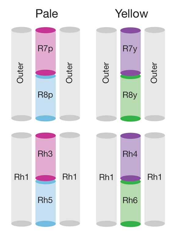
A photoreceptor cell is a specialized type of neuroepithelial cell found in the retina that is capable of visual phototransduction. These cells convert light (visible electromagnetic radiation) into signals that can stimulate biological processes, primarily by photoreceptor proteins absorbing photons and triggering a change in the cell's membrane potential. In mammalian eyes, three known types exist: rods, cones, and intrinsically photosensitive retinal ganglion cells.
- types
- Rods, cones, intrinsically photosensitive retinal ganglion cells
- location
- Retina (outermost layer for rods and cones)
- key function
- Convert light into neural signals via phototransduction
- color vision basis
- Ratios of responses from three cone classes (L, M, S)
Lore & Background
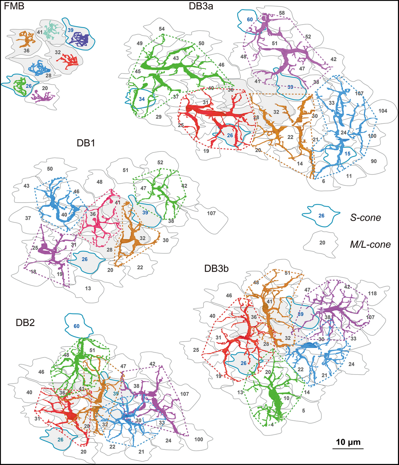
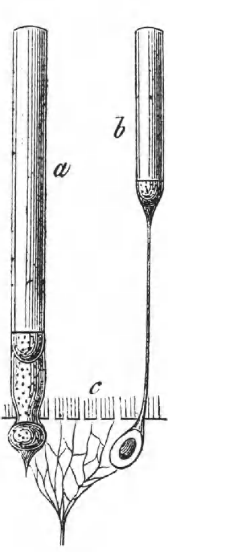
Photoreceptor cells are found on the outermost layer of the retina, with rods and cones sharing a basic structure: axon terminal, cell body, inner segment (rich in mitochondria), and outer segment (modified cilia containing opsin-filled disks). The outer segment absorbs light, while the inner segment provides ATP for the sodium-potassium pump. Rods primarily mediate scotopic vision (dim conditions), cones mediate photopic vision (bright conditions), and intrinsically photosensitive retinal ganglion cells, discovered in the 1990s, contribute to circadian rhythm entrainment and the pupillary reflex rather than sight directly. Each photoreceptor absorbs light according to its spectral sensitivity, determined by expressed photoreceptor proteins. The principle of univariance states that a photoreceptor's output is proportional only to the number of photons absorbed, not wavelength, so color vision arises from comparing ratios of the three cone responses. In the retinal mosaic, the fovea contains only cones and provides highest visual acuity, while rods and cones are intermingled elsewhere. No photoreceptors exist at the blind spot. The distribution of cone classes is nonhomogeneous, with no S-cones in the fovea and varying L-to-M cone ratios among individuals. Species vary in rod-to-cone ratios and number of cone classes, from monochromats to pentachromats.
Reader's Guide
Photoreceptor cells are fundamental to vision and non-visual light responses. Their ability to convert photons into electrical signals through the phototransduction cascade enables sight, with rods and cones forming the basis of image formation. The discovery of intrinsically photosensitive retinal ganglion cells expanded understanding of how light regulates circadian rhythms and pupillary reflexes. The principle of univariance explains why color perception requires multiple cone types, and the retinal mosaic's organization—especially the fovea's cone-only region—underpins high-acuity vision. The phototransduction cascade, involving opsin, transducin, and cGMP, illustrates a sophisticated amplification mechanism where a single photon can trigger a measurable cellular response. This knowledge has implications for treating retinal diseases and understanding evolutionary adaptations in diurnal versus nocturnal species.
Did You Know?
- Photoreceptors become hyperpolarized when stimulated, unlike most sensory cells, and release glutamate continuously in the dark.
- The outer segment of rods and cones contains disks filled with opsin, the molecule that absorbs photons.
- Melanopsin, found in intrinsically photosensitive retinal ganglion cells, functionally resembles invertebrate opsins.
Anatomy and Distribution in the Retina
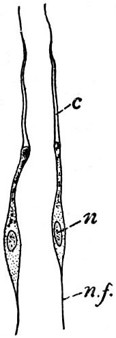
They share a fundamental architecture with cones: a synaptic terminal, an inner segment housing the nucleus and organelles, and an outer segment packed with stacked, opsin-containing disks that face the retinal pigment epithelium. A cilium bridges the inner and outer segments, lining the distal portion. The outer segment, oriented toward the back of the eye, holds the light-absorbing materials, while the synaptic terminal forms connections with bipolar or horizontal cells. Rods dominate the human retina in numbers—roughly 92 million compared to about 4.6 million cones—and they cluster heavily along the outer margins of the retina, making peripheral vision their primary domain. Despite this numerical advantage, rods contribute almost nothing to color perception, which explains why hues fade dramatically under dim lighting conditions.
The Molecular Cascade of Light Detection
When a photon strikes a rod cell, it triggers a chain reaction known as visual phototransduction. The light-sensitive pigment rhodopsin—composed of the membrane protein opsin covalently bound to a retinal molecule derived from vitamin A—undergoes a structural shift from 11-cis-retinal to all-trans-retinal. This conformational change boosts rhodopsin's affinity for transducin, a G protein whose alpha subunit swaps GDP for GTP and then detaches from its beta-gamma partners. The freed alpha subunit binds to cGMP phosphodiesterase, neutralizing the inhibitory gamma subunits and activating the enzyme. PDE then hydrolyzes cGMP into 5'-GMP, causing the cGMP-gated ion channels, primarily sodium with some calcium, to close. The loss of positive ion influx hyperpolarizes the cell, halting the spontaneous release of glutamate that occurs in darkness. This reversal of the resting depolarized state constitutes the fundamental electrical signal that light has been detected.
Sensitivity, Amplification, and the Vitamin A Connection
Rods achieve extraordinary light sensitivity through a powerful amplification cascade. A single activated rhodopsin molecule can recruit hundreds of transducin molecules, each of which in turn activates a phosphodiesterase enzyme capable of breaking down more than a thousand cGMP molecules per second. This multiplicative effect means even a handful of photons can produce a substantial electrical response, making rods almost entirely responsible for night vision. The stacked-disc architecture of the outer segment further enhances detection efficiency by packing a high density of opsin into a compact volume. However, this sensitivity comes with a trade-off: rods play virtually no role in color discrimination, which is why the world appears washed out and monochromatic in low light. Because retinal is synthesized from vitamin A, a dietary deficiency depletes the pigment supply in rod cells, reducing their capacity to respond in darkness and ultimately producing night-blindness—a condition that underscores the intimate link between nutrition and visual function.
Returning to Rest: Negative Feedback Mechanisms
After a flash of light, a rod cell must rapidly reset to its dark-adapted resting state to remain responsive to subsequent stimuli. Three inhibitory mechanisms work in concert to shut down the phototransduction cascade. First, rhodopsin kinase phosphorylates the cytosolic tail of activated rhodopsin at multiple serine residues, partially blocking further transducin activation. The inhibitory protein arrestin then binds to these phosphorylated rhodopsin molecules, fully silencing their activity. Second, an RGS protein acting as a GTPase-activating protein accelerates the hydrolysis of GTP back to GDP on the transducin alpha subunit, driving the G protein into its inactive off conformation. Third, as cGMP levels fall, the previously open cGMP-sensitive ion channels close, reducing the influx of positive ions and restoring the cell's membrane potential. Together, these layered feedback loops ensure that the rod cell recovers quickly and is ready to detect the next photon.
Gallery
Frequently Asked Questions
Who is Photoreceptor cell?
Photoreceptor cell is a specialized neuroepithelial cell stationed in the retina that acts as the eye's dedicated light-detection unit. It is a distinct member of the retinal neuronal cast, uniquely equipped to kick off the visual phototransduction cascade.
What are Photoreceptor cell's powers/role?
Its signature ability is converting incoming visible photons into electrical signals by using photoreceptor proteins to shift the cell's membrane potential. This phototransduction step is the very first link in turning raw light into information the brain can interpret.
How does Photoreceptor cell's story end?
In the mammalian retina, photoreceptor outer segments are continuously shed and phagocytosed by retinal pigment epithelium cells, while the cell body itself persists for extended periods. When the supporting RPE or the cell's internal machinery deteriorates, the photoreceptor degenerates and is lost, a process at the heart of conditions like retinitis pigmentosa.
What are Photoreceptor cell's main subtypes or ally factions?
Mammalian retinas feature three recognized photoreceptor lineages: rods for low-light vision, cones (divided into L, M, and S classes) for color and acuity, and intrinsically photosensitive retinal ganglion cells for non-image-forming light responses. The relative ratios of L, M, and S cone signals are what the brain ultimately reads as hue.
More in Cell And Molecular Biology 25-36
Elsewhere in the Cell And Molecular Biology universe
Spotted an error? Know more?
This is a living reference — every entry is fact-audited, and reader corrections feed straight into our audit queue. Suggest an edit · See this site's audit record