Mast cell
Ancient immune sentinels storing histamine and heparin.

John Jennings · CC BY-SA 2.0
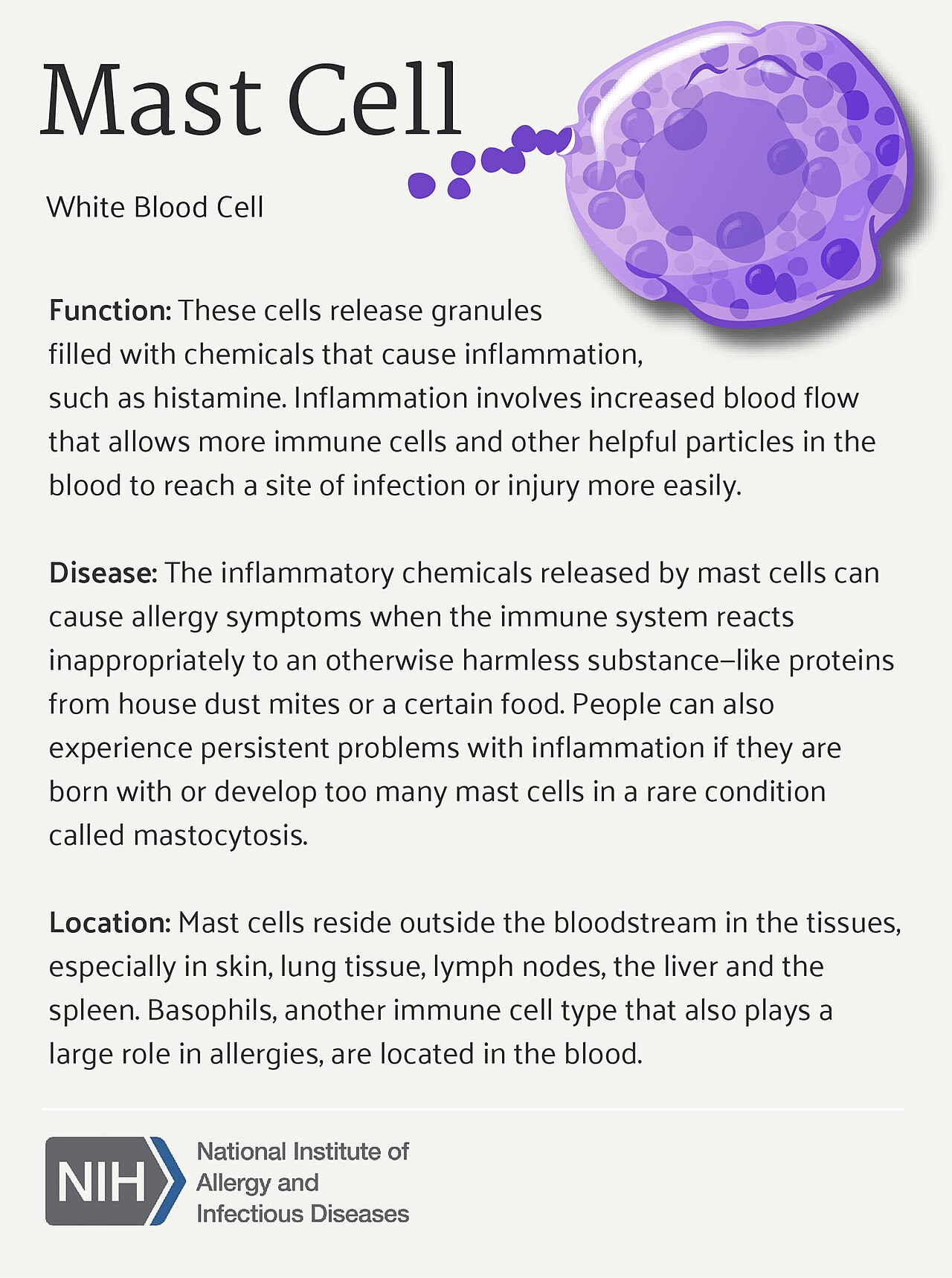
Mast cells, also known as mastocytes or labrocytes, are resident cells that develop and live in connective or mucosal tissue. They contain many small secretory granules for the storage and release of histamine, heparin, and other mediators. Derived from myeloid progenitor cells, mast cells are granulocytes, a type of white blood cell, and part of the immune and neuroimmune systems. They act as sentinels, detecting signals that indicate the presence of parasites, pathogens, and other possible dangers, and modulate immune responses by releasing stored and newly synthesized mediators.
- cell type
- Granulocyte, white blood cell
- origin
- Myeloid progenitor cells
- key mediators
- Histamine, heparin
- known for
- Roles in allergy, anaphylaxis, atopic dermatitis
Lore & Background
They develop from circulating mast cell progenitors (MCps) that, once recruited to connective or mucosal tissue, specialize and become resident mast cells. Mature MCs exhibit context-specific effector properties related to tissue types and diseases, and are highly varied. Mast cells in different tissues, such as gut and skin, exhibit different physical, behavioral, and biochemical characteristics and functions. Mast cells may have dual methods of origin in the hematopoietic system. The original layered immune theory proposed that hematopoietic stem cells (HSCs) were the basis for such development, but subsequent research suggests that multiple waves of immune cells develop from hemogenic endothelial cells, independent of HSCs. In vertebrates, the earliest source of mast cells is the extraembryonic yolk sac. During embryonic development, mast cell progenitors form in a series of developmentally discrete waves. The first wave is derived from erythro-myeloid progenitors in the yolk sac, before HSCs emerge. In humans, the first yolk sac-derived MCs originate from mesodermal precursors in blood islands of the yolk sac, starting around three weeks into gestation. Sizeable populations of fetal-derived MCs persist in connective tissue into adulthood and appear to self-maintain mostly independent of bone marrow.
Reader's Guide
Mast cells are significant as ancient immune cells that play a protective role in defense and repair, including wound healing, angiogenesis, vascular permeability, and responses to bacteria, viruses, protozoa, prions, fungi, and venoms. They are best known for their roles in allergy, anaphylaxis, and atopic dermatitis, and may be involved in a variety of other diseases. Their development involves multiple waves from yolk sac and fetal liver progenitors, with debate over whether adult mast cells originate from hematopoietic stem cells or are largely independent. Mast cells are categorized by tissue location and protease content: in rodents, connective tissue mast cells and mucosal mast cells; in humans, MCT (tryptase), MCTC (tryptase, chymase, carboxypeptidase), and MCC (chymase only). Their heterogeneity reflects both developmental origin and microenvironment. The persistence of fetal-derived mast cells into adulthood suggests a self-maintaining population, challenging simple models of immune cell renewal.
Did You Know?
- In humans, the first yolk sac-derived mast cells originate around three weeks into gestation.
- Sizeable populations of fetal-derived mast cells persist in connective tissue into adulthood and appear to self-maintain mostly independent of bone marrow.
An Ancient Immune Lineage
This deep evolutionary heritage places them among the most ancient immune cell types known, predating the emergence of many vertebrate-specific defenses. Their presence in such early chordate ancestors suggests that the basic capacity to store and deploy chemical mediators in response to environmental threats is a foundational feature of animal immunity. In modern vertebrates, mast cells occupy a distinctive niche as resident cells embedded in connective and mucosal tissues rather than circulating freely. They are classified as granulocytes within the broader family of white blood cells, derived from myeloid progenitor lineages, and they participate in both classical immune surveillance and neuroimmune signaling. The cell goes by several names in the literature—mastocyte, labrocyte—reflecting the layered history of its identification.
The Hematopoietic Debate
The developmental origins of mast cells remain one of the most actively debated questions in hematopoietic biology. In the classical model, hematopoietic stem cells give rise to multipotent progenitors, then common myeloid progenitors, then granulocyte-monocyte progenitors, from which mast cells and basophils differentiate. Research since the late 1980s has complicated this picture. Subsequent work suggests the earliest mast cell progenitors in vertebrates originate in the extraembryonic yolk sac, before hematopoietic stem cells even emerge. In mouse models, the first wave derives from erythro-myeloid progenitors in the yolk sac, with progenitors appearing around embryonic day 7.5 and maturing in the fetal liver by day 11. In humans, yolk sac-derived mast cells begin forming around three weeks of gestation. A critical unresolved question is whether adult mast cells in connective tissue are largely fetal-derived and self-maintaining, or whether they continue to be replenished from bone marrow stem cells. Sizeable populations of fetal-origin mast cells persist into adulthood, appearing to sustain themselves largely independent of bone marrow, yet myeloid precursors of mast cell lineage are found in bone marrow though mature cells are absent there.
Sentinel Function and Tissue Defense
Mast cells function as embedded sentinels within connective and mucosal tissues, constantly monitoring their surroundings for signals that indicate the presence of parasites, pathogens, or other potential dangers. Rather than operating with a single fixed response, they modulate their immune output according to the specific stimuli they detect. This flexibility is achieved through two complementary mechanisms: releasing pre-stored mediators from their numerous secretory granules and synthesizing new mediators on demand. The granules themselves are packed with histamine, heparin, and a range of other chemical agents that can be deployed rapidly when a threat is identified. Beyond their well-known role in allergic reactions, mast cells play a significant protective part in tissue defense and repair. They contribute to wound healing, promote angiogenesis, regulate vascular permeability, and mount responses against a broad spectrum of invaders including bacteria, viruses, protozoa, prions, fungi, and animal venoms. Their effector properties are context-specific, meaning that mast cells residing in the gut will behave differently from those in the skin, reflecting the distinct physical, biochemical, and functional demands of each tissue environment.
Clinical Significance and Disease Associations
While mast cells are essential guardians of tissue integrity, their very potency makes them central players in several pathological conditions. They are best known for their prominent roles in allergy, anaphylaxis, and atopic dermatitis, conditions in which their mediator release—particularly histamine—drives the hallmark symptoms of inflammation, swelling, and vascular changes. The same granule contents that protect against parasites and pathogens can, when deployed inappropriately or excessively, produce the dramatic and potentially life-threatening reactions seen in severe allergic episodes. Beyond these classic associations, mast cells may be implicated in a variety of other diseases, though the precise mechanisms and extent of their involvement in these additional conditions remain areas of active investigation. Their distribution is remarkably widespread: after maturing in their resident tissues, mast cells are found throughout the body, including in the brain, heart, lung, peritoneal cavity, skin, and spleen. This broad distribution means that their dysregulated activity could theoretically affect multiple organ systems. Understanding the context-specific behavior of mast cells in different tissues is therefore critical for developing targeted therapeutic strategies that can harness their protective functions while mitigating their pathological contributions.
Gallery



Frequently Asked Questions
Who is Mast cell?
Mast cell (a.k.a. mastocyte or labrocyte) is a tissue-resident sentinel that permanently stations itself in connective and mucosal tissues. It is classified as a granulocyte and a white blood cell, serving as a key operative in both the immune and neuroimmune systems.
What are Mast cell's powers/role?
Mast cell functions as a first-responder watchdog, detecting danger signals from parasites, pathogens, and other threats, then deploying its granule-packed arsenal—chiefly histamine and heparin—to shape the surrounding immune response. It can also whip up freshly synthesized mediators on the fly when the situation escalates.
Where does Mast cell come from?
Mast cell is generated from myeloid progenitor cells, anchoring it squarely in the myeloid branch of leukocyte development. Unlike circulating leukocytes, it migrates into tissue and remains a long-term resident rather than a transient visitor.
What is Mast cell most famous for?
Mast cell is the star of the allergy and anaphylaxis arcs, with a central role in atopic dermatitis as well. Its dense secretory granules loaded with histamine and heparin are what drive those dramatic, high-intensity immune reactions fans keep referencing.
More in Cell And Molecular Biology 1-24
Elsewhere in the Cell And Molecular Biology universe
Spotted an error? Know more?
This is a living reference — every entry is fact-audited, and reader corrections feed straight into our audit queue. Suggest an edit · See this site's audit record